Wound Healing Medical
Far Infrared Therapy in Wound Healing
A Mechanistic and Clinical Review, with Explicit Positioning on Chronic Wounds, Neuropathy, and Ischemia
ABSTRACT
Background: Wound healing proceeds through hemostasis, inflammation, proliferation, and remodeling. Chronic wounds, including diabetic foot, venous, and pressure ulcers, stall in a persistent inflammatory state driven by impaired perfusion, oxidative stress, infection, and repeated trauma. Standard care centers on debridement, moist wound management, infection control, offloading or compression, and correction of underlying disease.
Evidence: In animal models, FIR accelerated wound closure and collagen deposition without raising skin temperature (Toyokawa et al. 2003), sped diabetic wound healing in a dose-dependent manner with increased angiogenic signaling (Chen et al. 2021), and outperformed a heat-matched control in diabetic wounds while shifting macrophages toward a reparative phenotype (Jian et al. 2026). Human data are limited to perfusion studies (Kraaij et al. 2024) and one controlled diabetic foot ulcer trial of a broad-spectrum infrared lamp (Hakim et al. 2016).
Positioning: FIR is a promising investigational adjunct with consistent preclinical evidence and a plausible, partly non-thermal mechanism. It is not a substitute for professional wound care. Heat must not be applied to insensate or ischemic tissue, and no human trial has established FIR for chronic wound healing.
1. Introduction
Acute wounds in healthy individuals usually heal predictably. Chronic wounds do not. Diabetic foot ulcers, venous leg ulcers, and pressure injuries affect millions of people, carry high rates of infection and amputation, and consume a large share of healthcare resources. Their common features include poor tissue perfusion, persistent inflammation dominated by pro-inflammatory (M1) macrophages, excess oxidative stress, impaired angiogenesis, and reduced fibroblast activity.
Interest in far infrared (FIR) for wound healing stems from two observations: FIR increases skin microcirculation (Yu et al. 2006; Kraaij et al. 2024), and several animal studies report effects that persist when temperature is controlled, suggesting a wavelength-specific biological action (Toyokawa et al. 2003; Jian et al. 2026). This review evaluates that evidence and defines the boundaries for its use.
2. Biology of Healing and Its Failure
- Hemostasis (minutes to hours): Clot formation and release of growth factors.
- Inflammation (days): Neutrophils and macrophages clear debris and pathogens; a timely switch from M1 to reparative M2 macrophages is required for progression.
- Proliferation (days to weeks): Fibroblasts deposit collagen, endothelial cells form new vessels (angiogenesis), and keratinocytes re-epithelialize the surface.
- Remodeling (weeks to months): Collagen matures and reorganizes, increasing tensile strength.
- Chronic wound failure: Ischemia, hyperglycemia, oxidative stress, infection, and pressure or edema trap the wound in inflammation.
3. Far Infrared Biophysics and Delivery Formats
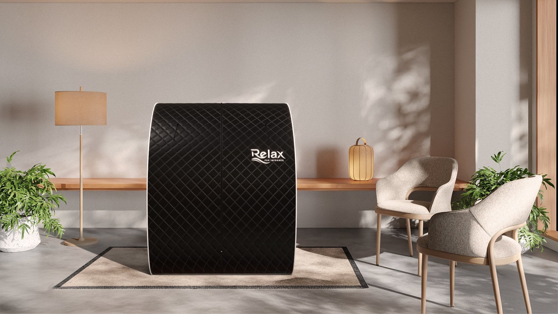
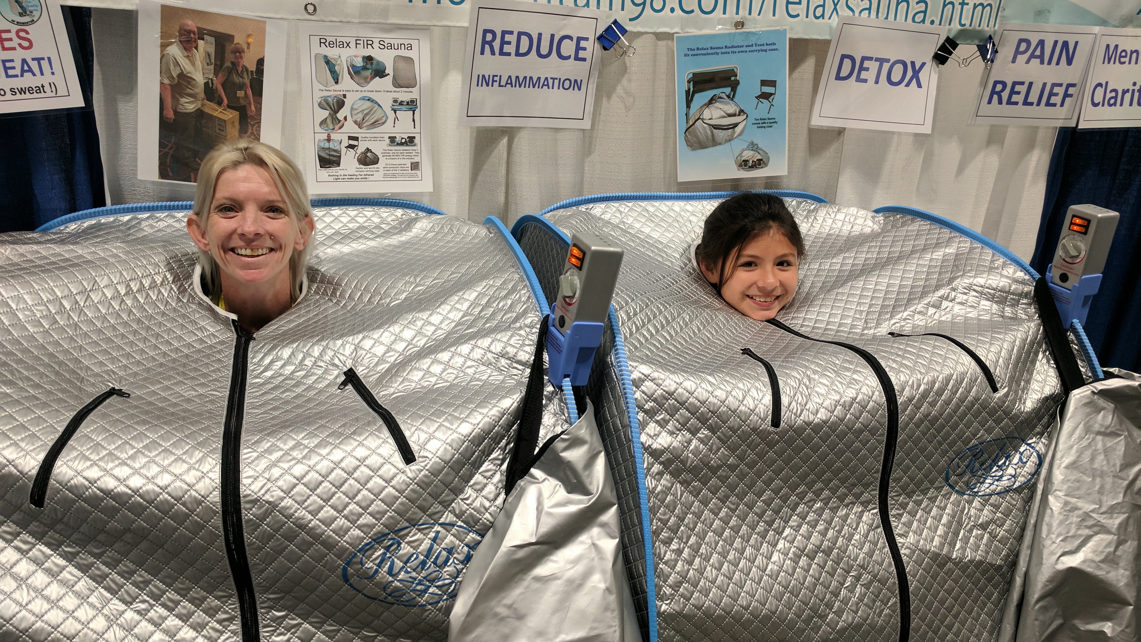
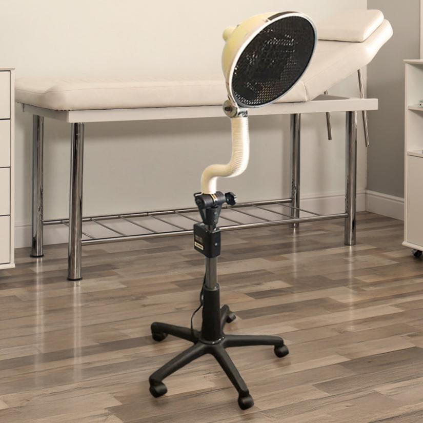
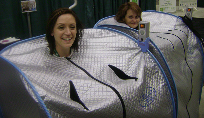
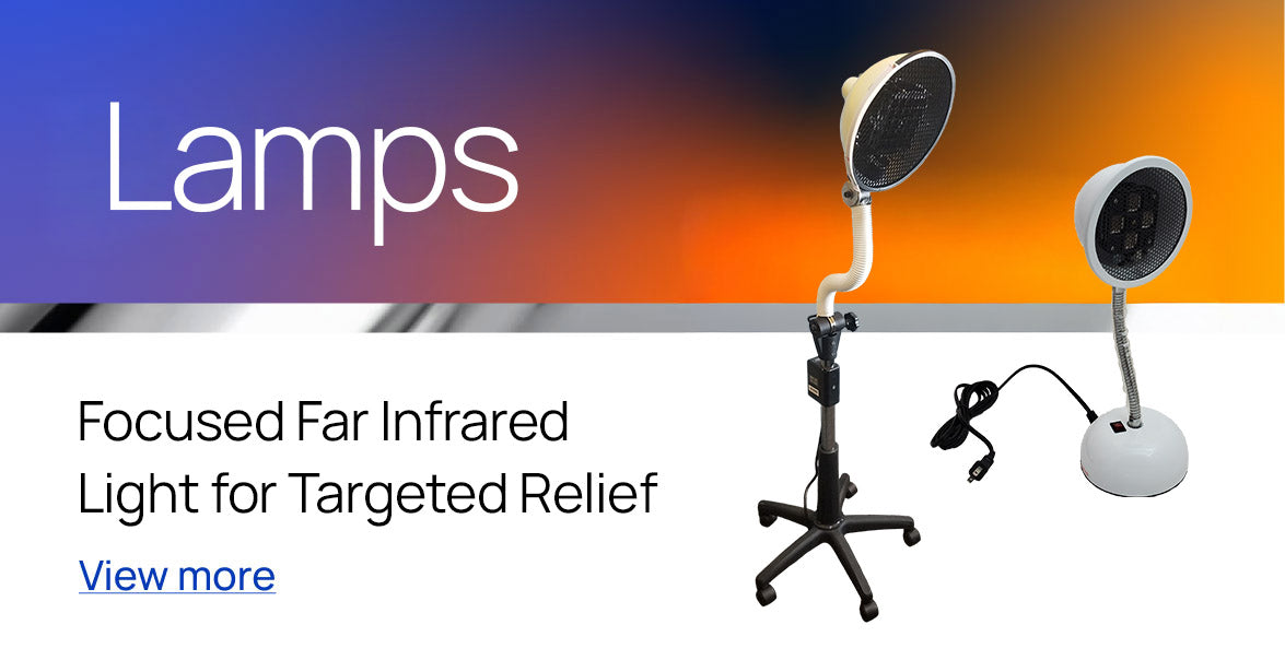
Far infrared radiation (approximately 4–14 µm) is absorbed predominantly in the superficial skin. Animal wound studies have used both very low-intensity emitters positioned at a distance (0.13 mW/cm² at 20 cm) (Chen et al. 2021) and heated emitters (Jian et al. 2026). Passive emitters, including mineral topicals and carbon or graphene textiles, provide low-intensity, non-heating exposure. Whole-body FIR, such as the Relax Sauna (PTC semiconductor emitters, 95–99% emissivity, 9.4 µm peak, head-out design), produces systemic warming and sweating and is not appropriate as a wound treatment while wounds are open.
4. Clinical Evidence
4.1 Acute wounds (animal)
In rats with full-thickness skin wounds, FIR accelerated wound closure by day 7 compared with both normal and temperature-matched controls, increased collagen fiber area from 26.5% to 56.8%, and increased fibroblast infiltration and TGF-β1 expression (Toyokawa et al. 2003). Skin temperature and blood flow did not change, supporting a non-thermal mechanism.
4.2 Diabetic wounds (animal)
In streptozotocin-diabetic rats with large full-thickness wounds, a low-intensity FIR emitter (5–12 µm, peak 8.2 µm) applied for 20 or 40 minutes three times weekly significantly accelerated healing compared with untreated diabetic controls (p<0.001), with 40-minute sessions healing faster than 20-minute sessions (5.8 vs 7.1 weeks) (Chen et al. 2021). FIR increased eNOS, VEGF, EGF, and Ki-67 and reduced inflammatory (CD45) and oxidative (8-OHdG) markers, with a dose-response pattern.
In a second diabetic rat model, a graphene FIR film and a metal heating film were both held at 41 °C, isolating the contribution of FIR emission from heat (Jian et al. 2026). At day 14, closure reached 83.9% with graphene FIR, 74.2% with the heat-matched control, and 66.8% in untreated diabetic wounds. Graphene FIR roughly halved wound TNF-α and IL-6, shifted macrophages from M1 toward M2, and increased Nrf2 and antioxidant gene expression.
4.3 Human evidence
In 50 patients with grade 1–2 diabetic foot ulcers, daily 20-minute infrared lamp treatment added to routine dressings for four weeks produced partial or full recovery in all treated patients versus 32% of controls, with worsening in 0% versus 20% (Hakim et al. 2016). The device was a tungsten infrared lamp with an unreported wavelength, likely weighted toward near infrared, and follow-up was short. In healthy adults, an FIR-emitting patch increased local skin perfusion and microcirculation (Kraaij et al. 2024).
Critical Appraisal & Limitations
The preclinical evidence is consistent across three independent rat studies and includes temperature-matched controls, which strengthens the case for a wavelength-specific effect. Human evidence is thin: no randomized trial has tested a defined FIR device on healing of chronic human wounds. The only clinical ulcer trial used a broad-spectrum lamp. Translation from rodent wounds, which heal partly by contraction, to human chronic wounds is uncertain.
4.4 Evidence Summary
| Study | Population / Model | Intervention | Key Finding | Tier |
|---|---|---|---|---|
| Toyokawa et al. 2003 | Full-thickness wounds, rat | FIR 5.6–25 µm (peak 8–12 µm) vs temperature-matched control | Faster closure by day 7; collagen area 56.8% vs 26.5%; more TGF-β1 fibroblasts; skin temp/blood flow unchanged | Direct FIR, animal (non-thermal) |
| Chen et al. 2021 | Diabetic rats, full-thickness wound, n=32 | FIR 5–12 µm (peak 8.2 µm), 0.13 mW/cm², 20 or 40 min 3×/wk | Faster healing vs controls (p<0.001); 40 min faster than 20 min (5.8 vs 7.1 wk); ↑eNOS, VEGF, EGF; ↓CD45, 8-OHdG | Direct FIR, animal (diabetic) |
| Jian et al. 2026 | Diabetic rats + cell models | Graphene FIR film vs metal film, both held at 41 °C | Day-14 closure 83.9% vs 74.2% vs 66.8% untreated; TNF-α and IL-6 roughly halved; M1→M2 shift; ↑Nrf2 | Direct FIR, animal (heat-matched) |
| Hakim et al. 2016 | Diabetic foot ulcer, n=50 | Infrared lamp (tungsten, wavelength not reported) + dressing vs dressing, 4 wk | Partial or full recovery 100% vs 32%; worsening 0% vs 20% | Adjacent IR, controlled trial |
| Yu et al. 2006 | Rat skin | FIR | Increased skin microcirculation | Mechanistic |
| Kraaij et al. 2024 | Healthy adults, n=20 | FIR-emitting patch, phase 1 RCT | Increased skin perfusion and microcirculation on multimodal imaging | Direct FIR, human mechanistic |
| Lin et al. 2008 | Endothelial cells and mice | FIR | HO-1 induction via NRF2/ARE; inhibited TNF-α-induced adhesion molecules | Mechanistic |
| Bagabir et al. 2025 | Human, heat-controlled 3-arm RCT | FIR vs heat vs control | FIR-specific NOS3, TXNRD1, HSP70 upregulation via NRF2 | Mechanistic |
5. Mechanistic Pathways
5.1 Angiogenesis & Perfusion
FIR increases skin microcirculation (Yu et al. 2006; Kraaij et al. 2024) and endothelial nitric oxide production (Park et al. 2013), and upregulated eNOS and VEGF in diabetic wounds (Chen et al. 2021). A heat-controlled human trial confirmed FIR-specific NOS3 upregulation (Bagabir et al. 2025).
5.2 Resolving Inflammation
FIR reduced TNF-α, IL-6, and inflammatory cell infiltration and promoted M1-to-M2 macrophage polarization in diabetic wounds (Chen et al. 2021; Jian et al. 2026). FIR also induces heme oxygenase-1 and restrains TNF-α-driven endothelial activation (Lin et al. 2008).
5.3 Oxidative Stress
Reduced 8-OHdG and increased Nrf2-regulated antioxidant genes in diabetic wounds (Chen et al. 2021; Jian et al. 2026) align with FIR-specific NRF2 activation in humans (Bagabir et al. 2025), linking wound effects to a pathway confirmed across tissues.
5.4 Fibroblast Activity & Collagen
FIR increased fibroblast infiltration, TGF-β1 expression, and collagen deposition (Toyokawa et al. 2003). Because TGF-β1 also drives fibrosis, effects on human scar quality cannot be assumed.
6. Explicit Positioning — What We Do Not Claim
- FIR is not a treatment for chronic wounds in humans. No randomized trial of a defined FIR device has shown improved healing of human chronic wounds.
- FIR does not replace debridement, infection management, offloading, compression, glycemic control, or vascular assessment.
- Heat must not be applied to insensate (neuropathic) or ischemic tissue, where it can cause burns or increase metabolic demand beyond blood supply.
- FIR does not treat wound infection and should never delay care for signs of infection.
- Animal findings, including graphene film results, should not be presented as human outcomes, and broad infrared lamp results should not be presented as FIR outcomes.
7. Open Question — What a Definitive Trial Would Need
- Population: Adults with non-ischemic diabetic foot ulcers (adequate perfusion confirmed by ankle-brachial or toe pressures) receiving standardized care including offloading.
- Arms: Standard care plus a defined, non-heating FIR emitter versus standard care plus sham emitter, with blinded wound assessment.
- Primary endpoint: Proportion of ulcers completely healed at 12 weeks.
- Secondary endpoints: Time to healing, wound area reduction, transcutaneous oxygen, infection, amputation, recurrence at 12 months, and adverse skin events.
- Dose characterization: Wavelength, irradiance, distance, skin temperature, and session duration recorded.
8. Clinical Guidance
Do Not
- Apply heat, heated emitters, or sauna exposure to feet or skin with peripheral neuropathy or reduced sensation; burns can occur without the patient feeling them.
- Apply local heat to wounds or limbs with significant arterial insufficiency, which can worsen ischemia.
- Use whole-body sauna while wounds are open; sweating, moisture, and shared surfaces risk maceration and contamination.
- Delay evaluation of spreading redness, warmth, swelling, odor, purulence, fever, or a wound that is not improving after two weeks.
- Apply topicals to open wounds unless specifically formulated and approved for that use.
Consider
- Referral to a wound care specialist for any chronic wound, and FIR only as an adjunct discussed with that team.
- Low-intensity, non-heating passive FIR near, but not on, intact skin surrounding a wound when sensation and perfusion are adequate.
- FIR around and over fully healed wounds and scars to support local circulation and comfort during remodeling.
- Whole-body FIR for general vascular health once wounds have closed.
9. Conclusion
Three independent animal studies show that FIR accelerates wound closure, angiogenesis, and resolution of inflammation, including in diabetic wounds, with effects that persist when heat is controlled. These findings align with FIR-specific NRF2 and nitric oxide signaling confirmed in humans. Human wound evidence remains limited to perfusion data and a single trial of a broad-spectrum infrared lamp. FIR is best positioned as a promising investigational adjunct for wound care, used only alongside professional management and never as heat on insensate or ischemic tissue, while well-designed human trials are undertaken.
At a Glance
- Preclinical Evidence: Consistent across 3 rat studies; accelerates closure & collagen
- Primary Mechanisms: eNOS/VEGF angiogenesis, macrophage M1→M2 shift, NRF2
- Human Evidence: Limited to perfusion data & 1 broad-spectrum lamp ulcer trial
- Key Cautions: Never apply heat to numb/neuropathic skin or ischemic limbs
- Role: Promising investigational adjunct in professional wound care
Clinical Precautions First
- Screen for peripheral neuropathy and loss of protective sensation
- Assess arterial perfusion (ABI, toe pressures) before thermal application
- Keep open wounds out of saunas; wait until epithelialization is complete
- Use passive products (Raylieve/HexTex) on healed scars/intact skin only
- Maintain standard wound care, offloading, and glycemic control
The Relax Sauna
Delivers pure 9.4 µm far-infrared wavelength energy with your head outside the cabin, supporting general systemic circulation and vascular health once wounds have fully closed.



Explore the Relax Far Infrared Line Up


Feel the Momentum